Understanding the Diagnostic Hysteroscopy Procedure: A Complete Guide to Women's Reproductive Health

Introduction to Diagnostic Hysteroscopy in Obstetrics & Gynecology
The diagnostic hysteroscopy procedure is a groundbreaking advancement in the field of gynecology, providing physicians with a minimally invasive method to visually assess the interior of the uterine cavity. As a vital diagnostic tool, this procedure empowers obstetricians and gynecologists to accurately identify and treat various uterine conditions, significantly improving women's reproductive health outcomes.
What Is a Diagnostic Hysteroscopy?
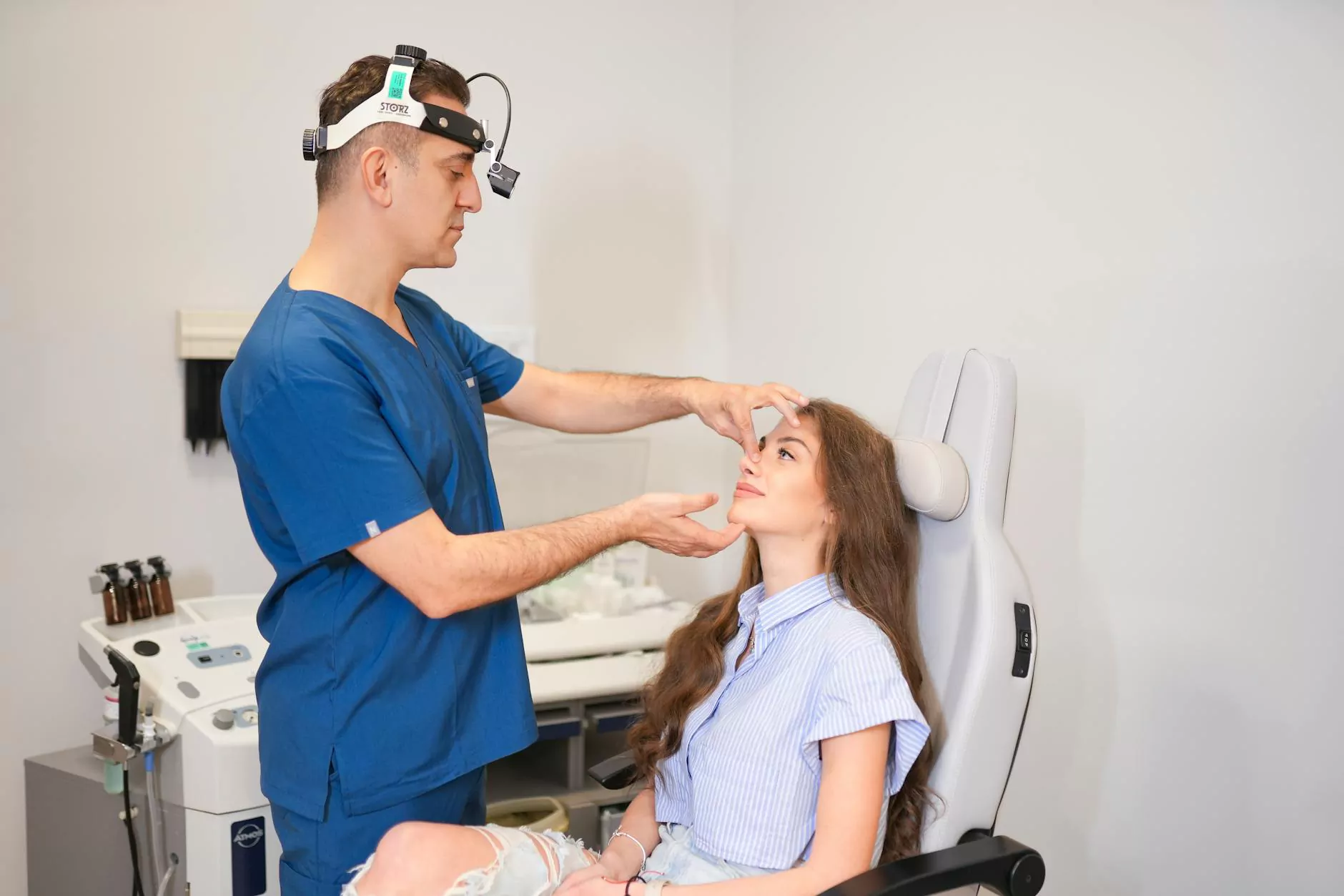
A diagnostic hysteroscopy involves the use of a specialized instrument called a hysteroscope—a thin, lighted tube that is gently inserted through the vagina and cervix into the uterus. Unlike traditional diagnostic practices, hysteroscopy provides real-time visual access to the uterine lining (endometrium), enabling direct examination rather than relying solely on ultrasound or other imaging techniques.
The Significance of Diagnostic Hysteroscopy in Women's Health
The role of diagnostic hysteroscopy procedure extends beyond simple visualization. It is instrumental in:
- Diagnosing uterine abnormalities: polyps, fibroids, septa, adhesions, and unusual tissue growths.
- Investigating causes of abnormal uterine bleeding which might include endometrial hyperplasia or polyps.
- Assessing infertility causes: identifying uterine factors that may hinder conception.
- Evaluating intrauterine pathology prior to treatment procedures.
Such precise diagnostics facilitate targeted treatments, minimize complications, and improve patient prognosis, making the diagnostic hysteroscopy procedure essential in modern obstetrics and gynecology.
Preparing for the Diagnostic Hysteroscopy Procedure
Preoperative Considerations
Before undergoing a diagnostic hysteroscopy procedure, patients should consult with an experienced obstetrician & gynecologist. Preparation may include:
- Complete medical history review and physical examination.
- Pelvic ultrasound to provide preliminary information about the uterine structure.
- Blood tests if necessary to evaluate overall health and detect any clotting disorders.
- Pregnancy test to confirm that the patient is not pregnant.
- Discussions about anesthesia options—local, regional, or general anesthesia—based on patient preference and clinical indications.
- Fasting or specific medication instructions if anesthesia is planned.
The Step-by-Step Process of Diagnostic Hysteroscopy Procedure
Step 1: Anesthesia Administration
Depending on the case and patient comfort, the procedure may be performed under local anesthesia (numbing the cervix), regional anesthesia (spinal or epidural), or general anesthesia. The goal is to ensure minimal discomfort during the examination.
Step 2: Insertion of the Hysteroscope
The obstetrician & gynecologist gently introduces a speculum into the vagina, then carefully dilates the cervix if necessary. The hysteroscope is then inserted through the dilated cervix into the uterine cavity. Gas (usually carbon dioxide) or fluid is used to distend the uterus, providing a clear view of the uterine walls.
Step 3: Visual Examination
Through high-resolution cameras attached to the hysteroscope, the physician inspects the uterine lining, detecting any anomalies such as polyps, fibroids, or adhesions. The real-time visualization allows immediate assessment, with the ability to take tissue biopsies if required.
Step 4: Tissue Sampling and Therapeutic Intervention
While primarily diagnostic, the diagnostic hysteroscopy procedure can be combined with targeted treatments like polypectomy, fibroid removal, or endometrial biopsy during the same session. This integrated approach enhances diagnostic accuracy and expedites patient care.
Step 5: Completion and Recovery
After thorough examination, the hysteroscope is gently withdrawn. Patients are monitored briefly post-procedure for any adverse reactions or discomfort. Many women experience minimal downtime and can resume normal activities quickly.
Benefits of Diagnostic Hysteroscopy in Modern Obstetric and Gynecological Practice
The adoption of diagnostic hysteroscopy in routine clinical practice offers numerous advantages:
- Minimal invasiveness: No large incisions or hospital stays required.
- High accuracy: Direct visualization allows precise diagnosis.
- Real-time biopsies: Immediate sampling for histopathological analysis.
- Combined diagnosis and treatment: Potential for same-session therapeutic procedures.
- Reduced patient discomfort and recovery time: Outpatient basis with fast return to daily activities.
Overall, this technique significantly enhances the scope and quality of gynecologic care, especially in complex cases involving infertility, abnormal bleeding, or suspected intrauterine pathology.
Recognizing When a Diagnostic Hysteroscopy Procedure Is Necessary
Obstetricians & gynecologists recommend diagnostic hysteroscopy in the following scenarios:
- Persistent abnormal uterine bleeding unresponsive to medical management.
- Infertility evaluations revealing intrauterine abnormalities.
- Recurrent miscarriage evaluations.
- Detection of uterine polyps, fibroids, or septa via ultrasound that need confirmation.
- Assessment of intrauterine adhesions or scar tissue, particularly after previous surgeries or infections.
- Preoperative assessment before operative hysteroscopic procedures.
Post-Procedure Care and Follow-Up
Post-procedure, patients might experience mild cramping, light bleeding, or spotting, which typically resolve quickly. Important post-care instructions include:
- Rest for the remainder of the day following anesthesia.
- Use of over-the-counter pain relievers if necessary.
- Avoiding intravaginal activities, such as tampon use or sexual intercourse, for a few days.
- Monitoring for signs of infection or excessive bleeding.
- Scheduling follow-up appointments to discuss findings and further treatment if needed.
Why Choose Expert Obstetricians & Gynecologists for Your Diagnostic Hysteroscopy?
Proper execution of the diagnostic hysteroscopy procedure demands not only advanced technology but also extensive clinical expertise. Leading clinics like drseckin.com provide highly skilled obstetricians and gynecologists specializing in minimally invasive procedures. Their dedicated team ensures patient safety, comfort, and accurate diagnosis, making them a trusted choice for women seeking reproductive health solutions.
Conclusion: Emphasizing the Importance of Diagnostic Hysteroscopy in Female Reproductive Health
The diagnostic hysteroscopy procedure continues to revolutionize gynecologic diagnosis by providing a detailed, minimally invasive means to explore and evaluate the uterine environment. Early detection and precise diagnosis of intrauterine abnormalities can drastically improve outcomes in fertility, menstrual health, and overall well-being. As a cornerstone of comprehensive women's health care, it remains an indispensable tool in the arsenal of modern obstetricians and gynecologists.
Take the Next Step Towards Better Reproductive Health
If you suspect uterine issues or require detailed evaluation of your reproductive health, consulting a specialized obstetrician & gynecologist who offers diagnostic hysteroscopy procedure is essential. Visit drseckin.com to learn more about expert care, advanced diagnostic options, and personalized treatment approaches designed to optimize your health and fertility.